Fig. 5.
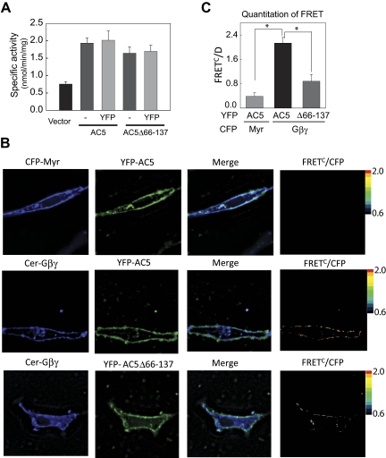
Cellular interaction of YFP-AC5 and Gβγ in COS7 cells by FRET. A, characterization of YFP-tagged proteins. Membranes from COS7 cells expressing vector, YFP-AC5, or YFPAC5Δ66–137 were stimulated with 1 μM GTPγS-Gαs. B, FRET analysis of AC5 and Gβ1γ2 in COS7 cells. Fluorescence microscopy images of COS7 cells transiently transfected with the indicated proteins were recorded using three different channels [1) donor, CFPex/CFPem; 2) FRET, CFPex/YFPem; and 3) acceptor, YFPex/YFPem]. A representative cell is shown for each combination of proteins (top, Myr-CFP and YFP-AC5; middle, Cer-Gβ1, Gγ2, and YFP-AC5; bottom, Cer-Gβ1, Gγ2, and YFP-AC5Δ66–137). Pseudocolor FRETC/D images were obtained using Slidebook software (Nikon) C, quantitative analysis of FRET by FC/D method (n = 4 using images from 18 to 20 cells, P < 0.01).

