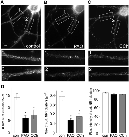
Fig. 4.
Blocking PIP2 synthesis or stimulating PIP2 hydrolysis decreases the surface NMDAR clusters on dendrites. A to C, immunocytochemical images of surface NR1 in cortical cultures treated without (control; A) or with PAO (10 μM; B) and carbachol (20 μM; C). Scale bars (A–C), 5 μm. Magnified versions of the boxed regions of dendrites (numbered 1 and 2) are shown beneath each image. D, quantitative analysis of surface NR1 clusters (density, size, and intensity) along dendrites under different treatments. ∗, p < 0.005, ANOVA, compared with control.

