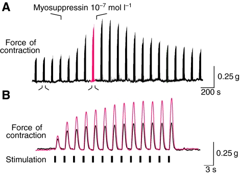
Fig. 10.
Myosuppressin perfused through the heart at 10−7 mol l−1 caused an increase in the amplitude of nerve-evoked contractions. (A) One of the anterolateral nerves was stimulated with 300 ms bursts of 60 Hz stimuli. Each burst caused a single contraction of the heart. Bursts were delivered in bouts of 13 bursts once every 2 min. (A) On a slow time base, each bout is compressed to form a single peak; the amplitude of these peaks more than doubled when 10−7 mol l−1 myosuppressin was perfused through the heart. (B) Faster time scale illustrates one bout of bursts in control (black, smaller contractions) and in myosuppressin (red, larger amplitude contractions). In this example, there was a substantial increase in facilitation in myosuppressin, but statistically, facilitation did not increase across multiple preparations.

