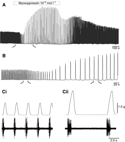
Fig. 3.
Myosuppressin decreased heart rate and had complex effects on amplitude when perfused through an isolated whole heart at 10−7 mol l−1. (A) Recording on a slow time base illustrates a typical response to myosuppressin: heart rate decreased, amplitude of contractions first decreased slightly (~10% in the example shown), then dramatically increased to over 200% of its original value. (B) Higher speed recording of initial response, taken from time demarcated in A by the black lines. (C) Higher speed recordings of heartbeat and associated neural activity, as recorded extracellularly on the anterior lateral nerve in control (Ci) and 4 min after the onset of myosuppressin perfusion (Cii), as demarcated in B. Cycle frequency decreased, contraction amplitude increased, and both burst and contraction duration increased in the presence of 10−7 mol l−1 myosuppressin.

