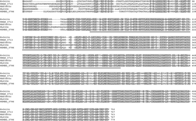
Fig. 1.

Alignment of mycobacterial protein kinase G orthologues. PknG sequences from M. tuberculosis H37Rv (Rv0410c, from the Pasteur Institute), M. marinum (MMAR_0713, from the Sanger Institute), M. avium paratuberculosis (MAP3893c, from TIGR), M. leprae (ML0304, from the Sanger Institute) and M. smegmatis (MSMEG_0786, from TIGR) were aligned using clustalw. The PknG orthologues of M. bovis and M. microti were omitted since they were identical to M. tuberculosis PknG. The M. smegmatis PknG coding sequence starts at the ATG that aligns to the start codon of M. tuberculosis PknG (see Fig. S1 online). Identical residues are shown in grey and the kinase domain is underlined.
