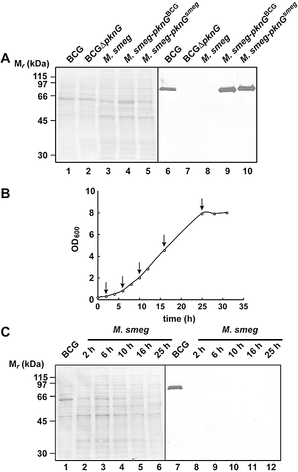
Fig. 4.

Expression of M. smegmatis PknG during in vitro growth. A. Lysates from M. bovis BCG (lane 1), M. bovis BCGΔpknG (lane 2), M. smegmatis (lane 3), M. smegmatis-pknGBCG (lane 4), M. smegmatis-pknGsmeg (lane 5) were separated on a 10% SDS-PAGE gel and immunoblotted using anti-PknGBCG antiserum (right). The total protein pattern was analysed by Ponceau Red staining (left). B and C. M. smegmatis was grown in 7H9-OADC medium until stationary phase. Bacterial samples were collected at different time points (arrows in B) and analysed for the presence of PknG by immunoblotting as under A (C). BCG lysate was loaded in parallel as a positive control (lane 1). Again, the total protein pattern was analysed by Ponceau Red staining (left). Results are representative data from one experiment.
