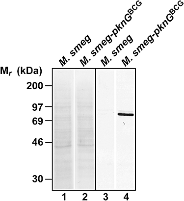
Fig. 5.

Expression of M. smegmatis PknG during macrophage infection. PknG immunoblot of macrophages infected for 16 h with M. smegmatis or M. smegmatis expressing PknGBCG. After infection, cells were harvested, homogenized and a post-nuclear supernatant was submitted to immunoblotting (lanes 3 and 4). The total protein pattern was analysed by Ponceau Red staining (lanes 1 and 2). Results are representative data from one experiment.
