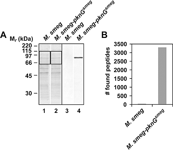
Fig. 6.

Selected ion monitoring analysis of M. smegmatis lysates. A. Lysates (10 μg) from M. smegmatis and M. smegmatis-pknGsmeg were separated on a 10% SDS-PAGE gel, followed by Coomassie Blue staining (lanes 1 and 2) or immunoblotting using anti-PknGBCG antiserum (lanes 3 and 4). B. Thirty micrograms of the same lysates as analysed under (A) were separated on a 10% SDS-PAGE gel and stained with Colloidal Blue. The region between 65 and 115 kDa was excised (indicated by the rectangles in A), digested with trypsin and the obtained peptides were identified using selected ion monitoring (see Experimental procedures). Representative data from two independent experiments are shown.
