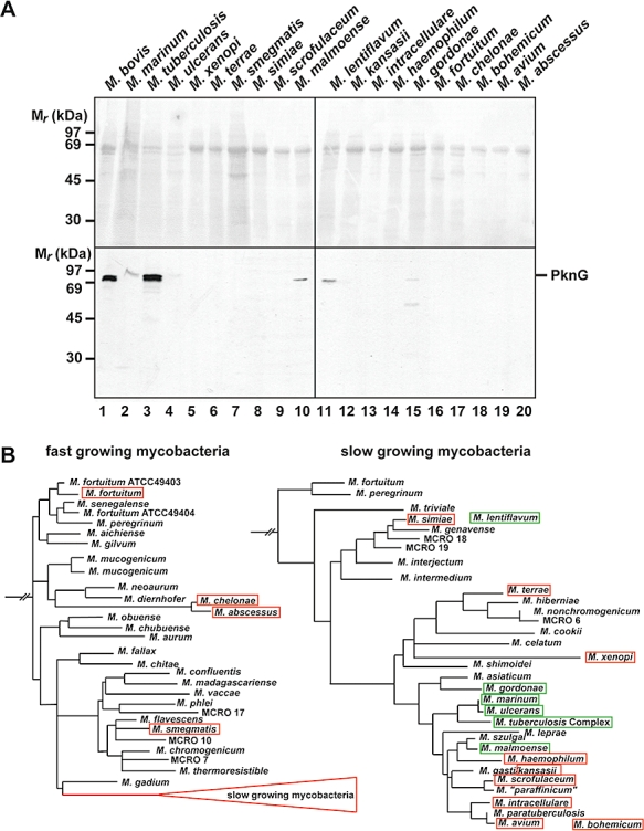
Fig. 9.

Analysis of pknG expression in a wide range of mycobacterial species. A. Lysates of various mycobacterial species were separated on a 10% SDS-PAGE gel and immunoblotted using anti-PknG antiserum (bottom). The total protein pattern was analysed by Ponceau Red staining (top). For sources and growth conditions of the mycobacterial species see Table S2. B. Phylogenic tree of mycobacteria based on 16S rRNA gene sequences. Species that show a PknG signal in (A) are boxed in green, the ones that do not show expression are boxed in red. Tree adapted from Springer et al. (1996).
