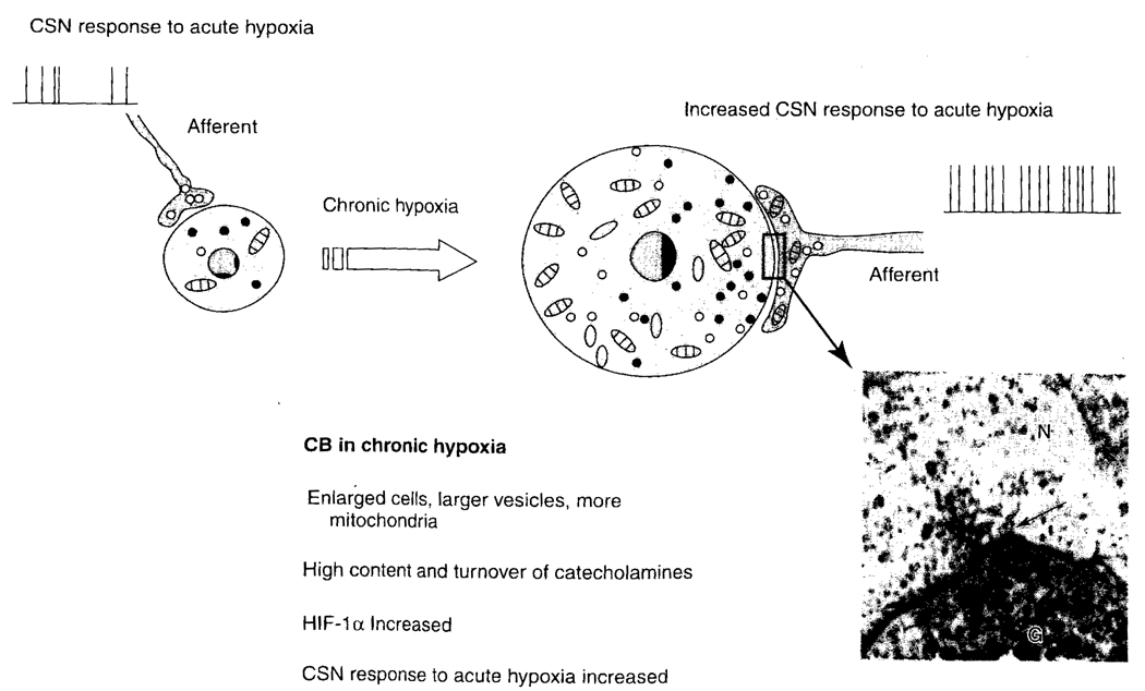
FIG. 5.
Schematic drawing of the changes in the CB during chronic hypoxia (modified after Joseph and Pequignot, 2003). Carotid bodies from animals subjected to chronic hypoxia have an increased response to acute hypoxia. N, nerve ending; G, glomus cell; the arrow indicates a synaptic connection.

