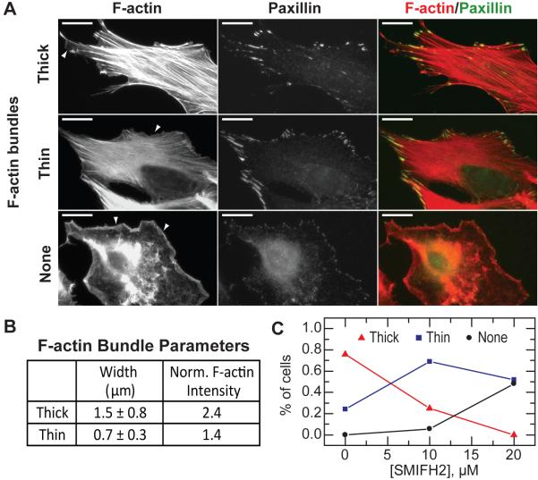
Figure 7. Effect of SMIFH2 on the Actin Cytoskeleton in NIH 3T3 Fibroblasts.
(A) Immunofluorescence of filamentous actin and paxillin in NIH 3T3 fibroblasts. Cells that remained spread after SMIFH2 treatment exhibited three distinct actin cytoskeleton organizations: thick F-actin bundles, thin F-actin bundles and no F-actin bundles. Lamellipodial actin is observed in all phenotypes, and indicated by arrows. Scale bar is 10 mm.
(B) F-actin bundles observed in (A) are characterized by their mean width and the maximum filamentous actin intensity normalized to the local background.
(C) Percentage of spread cells exhibiting different actin cytoskeleton phenotypes after 2 hr incubation with indicated concentrations of SMIFH2 (n > 75 cells for each concentration).

