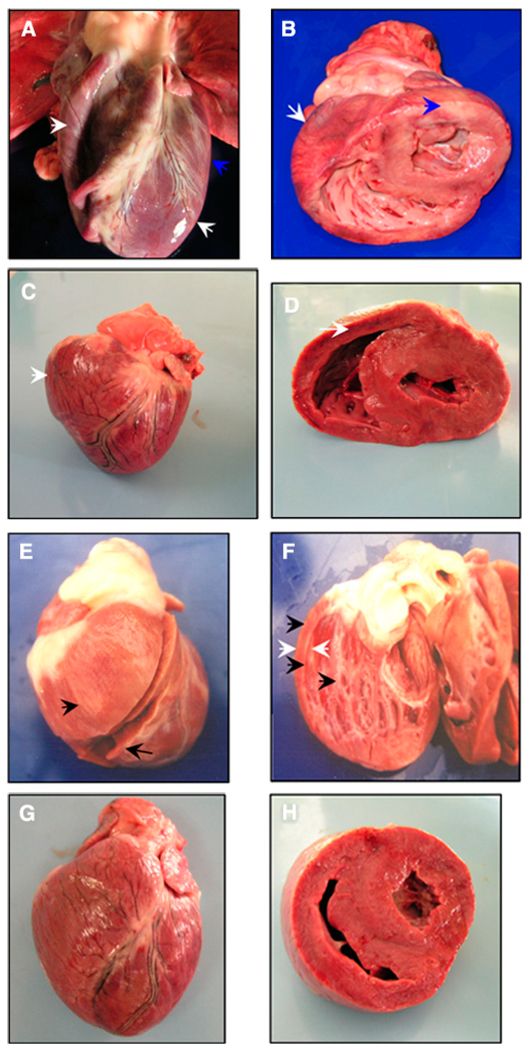
FIGURE 3.
Morphologic alterations of the Chagasic hearts from naturally and experimentally infected dogs. A and B, Heart morphology of a naturally infected dog with biventricular dilated cardiomyopathy (white arrows) and presence of pale striated epicardium (blue arrows). C and D, Heart from a Zumpahuacan-infected dog shows right-ventricle dilated cardiomyopathy and heart enlargement (white arrows). E and F, Heart from a Sylvio-X10–infected dog shows right-ventricle dilated cardiomyopathy with thin walls (white arrows) characterized by a rounded heart appearance and presence of whitish striates in epicardium, myocardium, and endocardium (black arrows). G and H, Heart from a healthy, non-infected dog. This figure appears in color at www.ajtmh.org.

