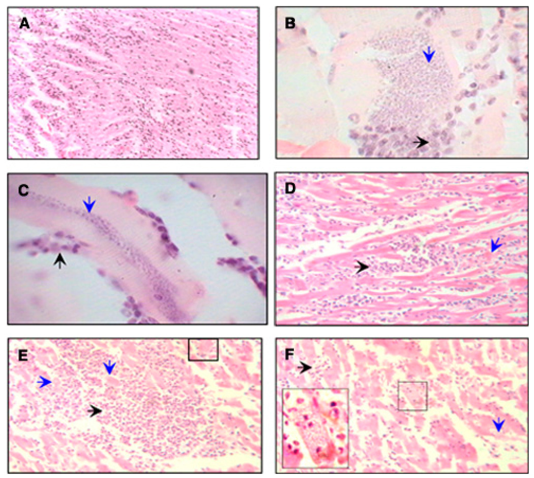
FIGURE 4.
Histopathological findings in naturally and experimentally infected dogs. Heart tissue sections (5 µm) were stained with hematoxylin and eosin. A, Note the infiltration of lymphoplasmacytic cells in myocardial section from a naturally infected dog (magnification: ×200). B and C, Transversal (B) and longitudinal sections (C) of muscular fiber from Zumpahuacan-infected dogs show the presence of large amastigote nests. In B, downward arrow shows the basophilic granular appearance of amastigotes inside of a muscular fiber and horizontal arrow shows the leukocyte inflammatory reaction against the infested fiber (×1,000). In C, the downward arrow shows the basophilic granular appearance of amastigotes in the middle of a muscular fiber, and the upward arrow shows the leukocyte inflammatory reaction against the infested fiber (magnification: ×1,000). D–F, Tissue sections from Sylvio-X10–infected dogs. D, Severe necrotic myocarditis characterized by leukocyte infiltration (horizontal arrow) with multifocal myofiber segmental necrosis (downward arrow) is evident. This infiltration corresponded to the macroscopic pale zones (whitish striates) observed in the heart. E, Dense focal interstitial leukocyte infiltration with severe multifocal myofiber segmental necrosis, loss of myocytes, and presence of a nest of amastigotes into a myofiber (small square). F, Moderate leukocyte infiltration (horizontal arrow) with multifocal myofiber segmental necrosis (downward arrow), and presence of a nest of amastigotes inside a myofiber (inset) (magnification: ×400). This figure appears in color at www.ajtmh.org.

