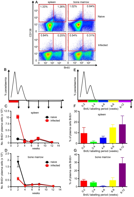
Figure 3. Plasma cells generated in the first 2 weeks of an acute P. chabaudi infection are not long-lived.
(A) Cartoon indicating the 2-week period of oral BrdU administration after infection of C57BL/6 mice with 105 P. chabaudi iRBC, and the subsequent timing of removal of spleens and bone marrow for the analysis shown in graphs B & C. Total numbers of BrdU-labelled CD138+ cells (gated as shown in Supplementary Figure S3) in spleen (B) and bone marrow (C) of P. chabaudi-infected mice. (D) Cartoon indicating the different 2- or 4-week time periods of oral BrdU administration following infection of C57BL/6 mice with 105 P. chabaudi iRBC for the analysis for graphs E & F. Spleens and bone marrows were removed and analysed after 12 weeks of infection. Percentage of CD138+ cells labelled with BrdU (gated as shown in Supplementary Figure S3) in spleens (E) and bone marrow (F) of infected mice at 12 weeks post-infection. The values shown are the mean number (B and C) or percentage of cells (E and F) from 5 individual mice, and the error bars represent the standard errors of the means.

