Figure 6.
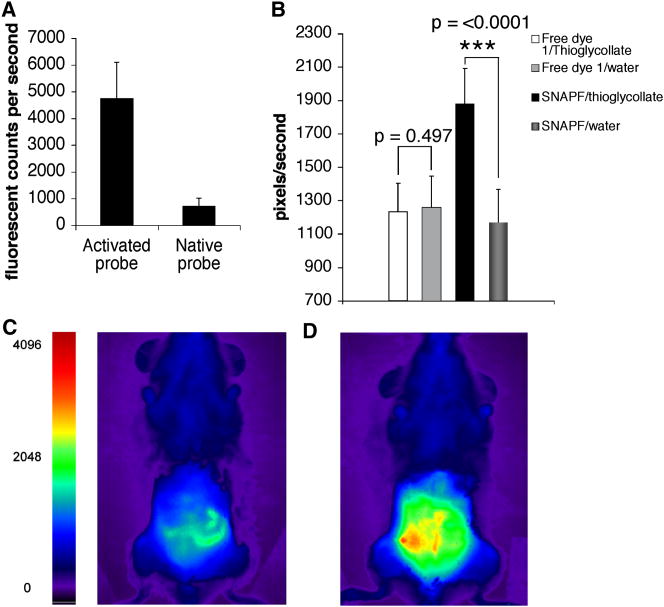
FRI imaging reveals SNAPF activation by HOCl in vivo. Comparison of FRI images of SNAPF alone or incubated with 20 μM HOCl shows an approximate 7-fold increase in SNAPF fluorescence in vitro using this imaging system (a). We then injected 50 nanomoles of SNAPF i.p into hMPO-Tg mice with thioglycollate-induced peritonitis and imaged the animals after one hour. There was a ~1.4-fold increase in peritnoneal fluorescence (b), presumably due to contact of SNAPF with HOCl generated by activated neutrophils and macrophages present. Fluorescence was ~1.6-fold higher post-SNAPF injection in mice injected with thioglycollate as opposed to water. c), FRI image of h-MPOtg mouse pre- SNAPF injection, d), the same animal post-injection of SNAPF. There was no visible fluorescence when SNAPF was injected into mice that had been injected with saline solution as opposed to thioglycollate, nor was there any increase in fluorescence of a wavelength-matched control dye, 1, when injected into mice with peritonitis compared to saline-injected animals (b) (n=4 per group).

