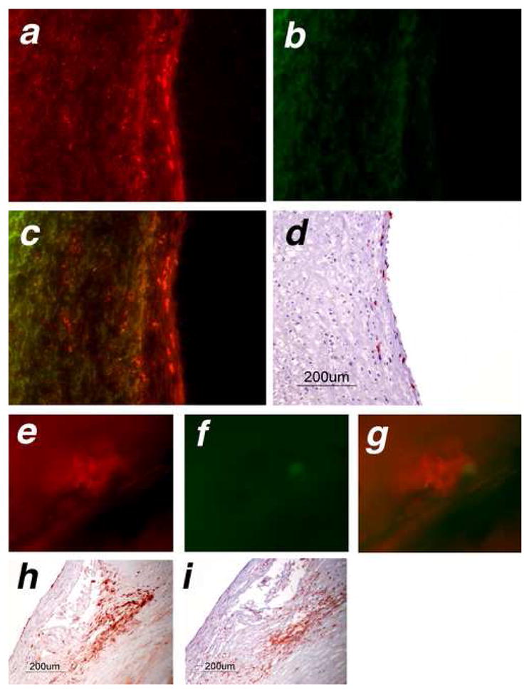
Figure 7.

SNAPF detects areas of HOCl generation in frozen sections of human atherosclerotic plaque, and is activated after incubation with fresh atheromatous carotid endarterectomies.. Frozen serial sections cut from atheromatous carotid endarterectomies (n=4) were incubated at 37°C for 1 hour with 10 μM SNAPF, or with buffer alone as a control, then viewed by fluorescence microscopy. Sections incubated with SNAPF were observed using an APC (a, colored in red) and a FITC (b, colored in green) filter set. Merged images (c) show that fluorescent areas using the APC filter set, which detects SNAPF, are distinct from areas that fluoresce using the FITC filter set (autofluorescence). The areas which are fluorescent using the APC filter set correspond to the location of MPO+ cells in the section (d).
Freshly excised human endarterectomies (n=4) were obtained from the operating theaters of the Brigham and Women’s Hospital, cut into thin slices and incubated with 10 μM SNAPF at 37°C for one hour. The slices were then viewed under a fluorescent microscope. Areas which were fluorescent when viewed using the APC filter set (e, pseudocolored in red) were not fluorescent with the FITC filter set (f, green) when exposed for the same amount of time, indicating that this was likely not autofluorescence. Merged images show distinct red colored areas (g). The slices contained MPO+ cells, established by immunohistochemical staining of adjacent tissue (h). MPO+ cells colocalised with cells, which stained positively for macrophages in adjacent sections (i).
