
Figure 2.
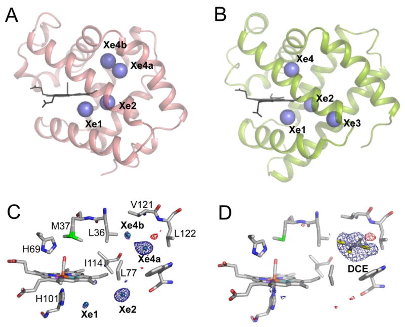
Xe and organic halide binding to HbI and Mb.
(A and B) Comparison of the Xe binding sites within HbI (A) and Mb (B; PDB file 1J52). The Xe1 and Xe2 sites from both globins are at nearly identical locations whereas Xe4a/Xe4b site in HbI is close to, but not at identical location as Xe4 in Mb. Xe3 in Mb is not present in HbI.
(C and D) A close up view of the Xe (C) and DCE (D) binding sites is superimposed upon the corresponding FoXe-FoNative or FoDCE-FoNative difference electron density map, contoured at ± 3σ. The DCE binding site is equivalent to the Xe4a binding site.