Fig. 2.

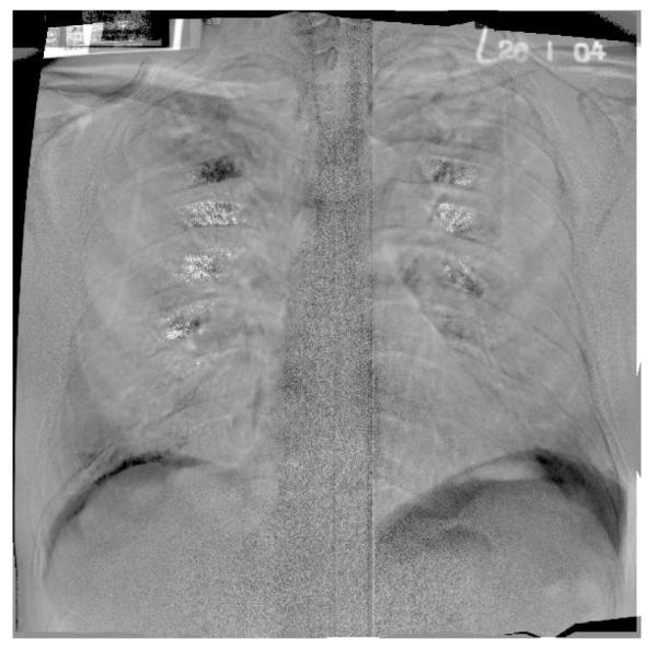
An example of a subtraction image using the entirety of both the first and second chest radiographs of a TB patient undergoing DOTS therapy. Note that some anatomical structures can still be identified.

An example of a subtraction image using the entirety of both the first and second chest radiographs of a TB patient undergoing DOTS therapy. Note that some anatomical structures can still be identified.