
Figure 2. Mutations in evi mimic abnormal synaptic phenotypes observed in wg mutants.
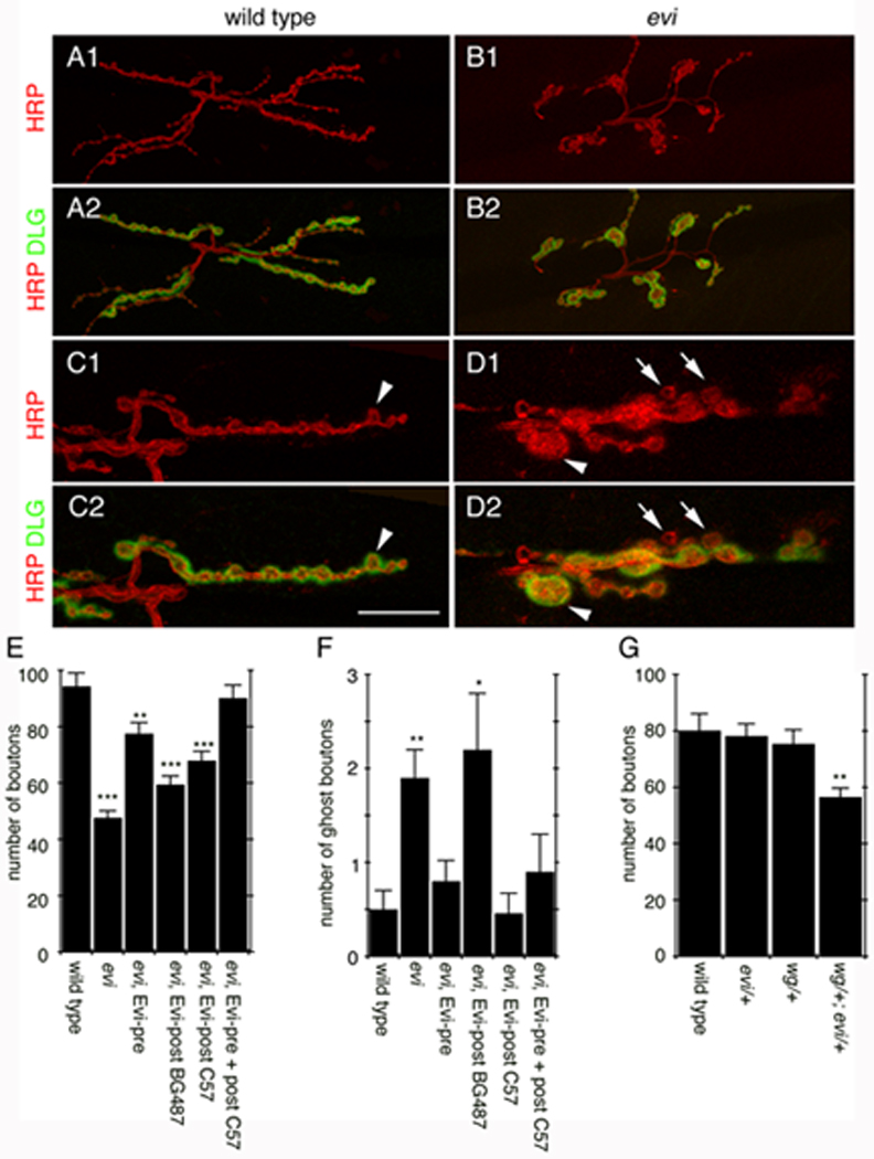
(A–D) Confocal images of NMJs labeled with anti-HRP and anti-DLG in (A, C) wild type, and (B, D) evi. (A, B) Projections of entire NMJs. (C, D) Single confocal slices of NMJ branches (arrowheads in C, D= abnormal boutons; arrows= ghost boutons). (E–G) Number of (E, G) boutons and (F) ghost boutons. Calibration bar is 30 µm for A, B and 13 µm for C, D.