
Figure 4. Evi is transferred from cell to cell and to the medium.
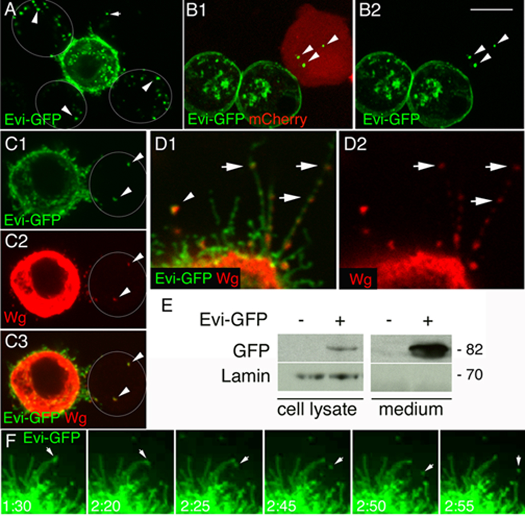
(A, B) Single confocal slices of S2 cells (A) either untransfected (outlined by white circles) or transfected with Evi-GFP and (B) either transfected with mCherry or Evi-GFP (arrowheads= Evi-negative cells; arrows= Evi in the media). (C) Evi-GFP and Wg are transferred together into an untransfected cell (arrowheads) (D) Wg localizes with Evi into punctuate structures within filopodia (arrows), as well as in the medium (arrowhead) (E) Western blot of lysates and media from Evi-GFP transfected S2 cells. (F) Time-lapse imaging of an S2 cell transfected with Evi-GFP showing the shedding of an Evi-GFP vesicle to the medium (arrows). Calibration bar is 3µm for panel 4D and 8 µm for the rest of the panels. Time points in 4F are in min.