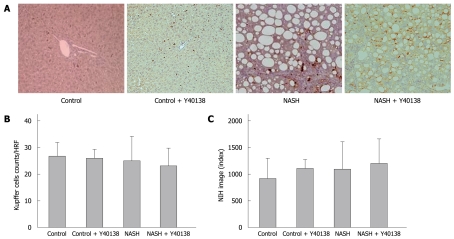
Figure 4.
Kupffer cell immunohistochemical staining, Kupffer cell number and semiquantification in the liver. A: Kupffer cell immunohistochemical staining (× 200). Brown-stained cells are positive; B: No significant differences were seen between groups in the stained cell counts per field; C: Quantitative analysis revealed no significant differences between groups (n = 5). HRF: High rise filed.

