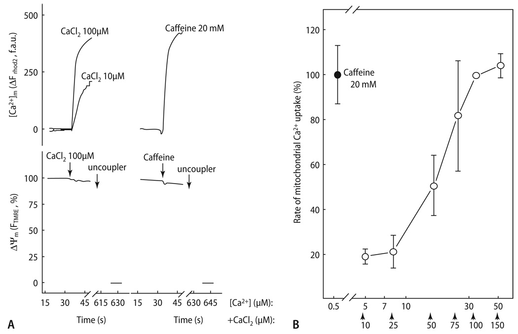
Fig. 7.
Full activation of mitochondrial Ca2+ uptake during RyR-mediated Ca2+ release. (A) Ca2+- and caffeine induced [Ca2+]m and Δψm responses were measured in permeabilized myotubes using rhod-2 and TMRM, respectively. Uncoupler (1 µM CCCP and 2.5 µg/ml oligomycin) was added as indicated. (B) The rate of mitochondrial Ca2+ uptake was measured at varying [Ca2+] obtained by the addition of CaCl2 in adherent rhod-2 loaded permeabilized cells. The added CaCl2 concentration values are indicated with arrows below the x axis, and the effective [Ca2+]c were calculated. To prevent Ca2+-induced Ca2+-release via RyR2, the cells were preincubated with thapsigargin to inhibit the SR Ca2+ ATPase and hence to deplete the SR prior to Ca2+ addition. Reproduced from Szalai et al. [191] with permission

