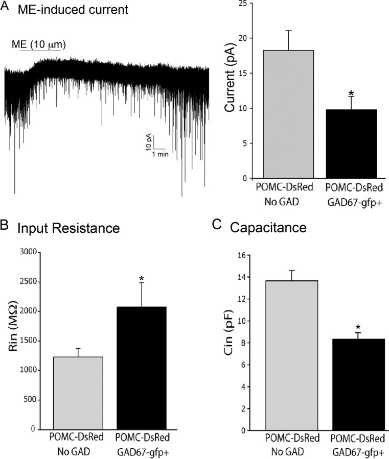
Figure 6.
Postsynaptic regulation and properties of GABAergic and non-GABAergic POMC neurons. A, Perfusion of ME (10 μm) caused an outward current in both GAD67–gfp-positive and GAD67–gfp-negative POMC neurons, although the average magnitude of the current was larger in POMC neurons without GAD (n = 8–10). The recording shown in A is a representative trace made in a GABAergic POMC neuron. B, Input resistance in GAD-negative (gray bars; n = 14) and GAD67–gfp-expressing (black bars; n = 15) POMC neurons. C, Capacitance measurements in GAD-negative (n = 12) and GAD67–gfp-expressing (n = 15) POMC neurons. *p < 0.05. Error bars represent SEM.

