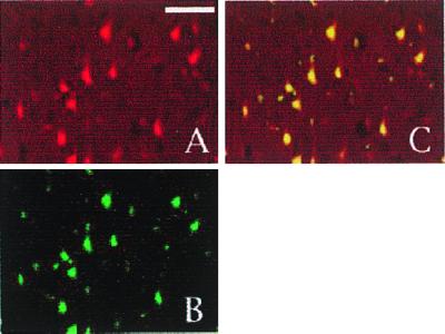
Figure 2.

Cyclin D1 immunoreactivity is localized to neurons in a confocal image of the core infarct region 24 h after reperfusion. (A and B) Sections were double labeled with cyclin D1 (A) and the neuronal marker NeuN (B). (C) The merged image. (The white bar represents 50 μm.)
