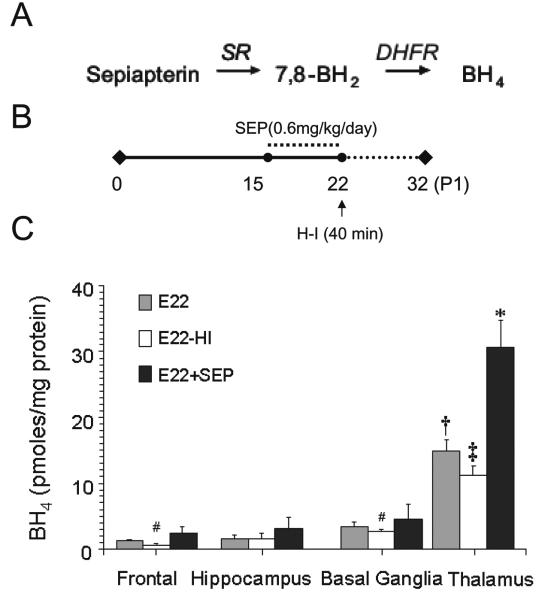
Figure 5.
Changes in BH4 concentrations in parts of the E22 fetal brain following H-I and sepiapterin supplementation. A, Stepwise conversion of sepiapterin to BH4 in reactions catalyzed by sepiapterin reductase (SR) and dihydrofolate reductase (DHFR). B, Sepiapterin treatment scheme. Treatment was initiated at E15 and finished at E22 covering from 48 to 70% term gestational period. Control dams received vehicle only. The E22 tissue from sham operated or H-I group was collected for BH4 measurements. C, Analysis of BH4 in different parts by HPLC-ECD. [E22-group]: Tissue was collected from fetuses retrieved from vehicle treated and sham operated dams at E22. [E22-H-I group]: Tissue was collected immediately after retrieving fetuses by hysterectomy from a dam that was subjected to sustained hypoxia for 40 min at E22. [E22+Sep group]: Tissue was collected from fetuses retrieved from dams receiving sepiapterin (0.6 mg/kg/day) during 5 days prior to sham-operation at E22. Values are mean±SE (n≥4). (†) p<0.05 with respect to other parts of the brain; (‡) p<0.05 with respect to E22 thalamus; (#) with respect to E22 frontal; (*) p<0.01 with respect to all experimental groups.

