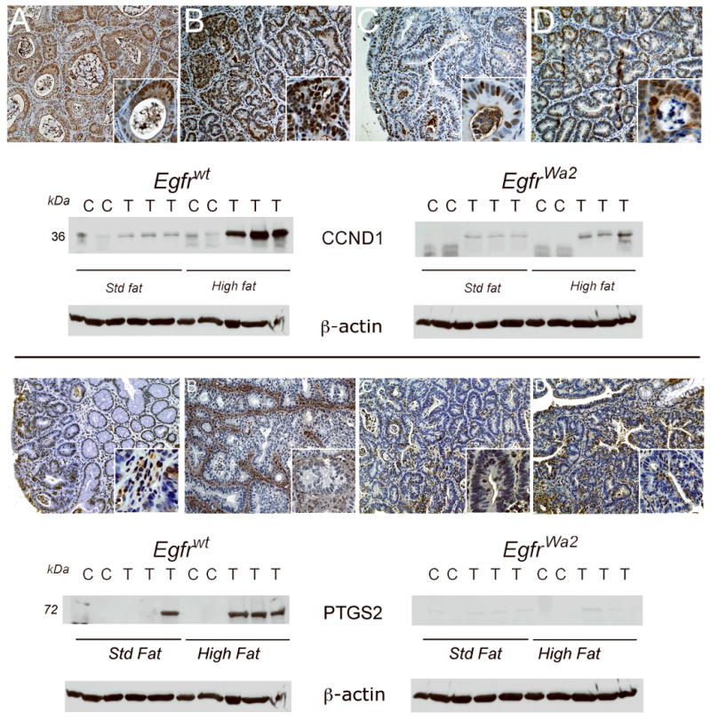
Fig. 3. CCND1 and PTGS2 expression levels in colonic tumors are controlled by Egfr genotype and diet.

Colonic tumors were immunostained and Western blotted as described in “Materials and Methods”. Shown are representative tumors from each group. Upper panel: CCND1 IHC. A. Egfrwt, std fat; B. Egfrwt, high fat; C. Egfrwa2, std fat; D. Egfrwa2, high fat. Images are 20× and insets 100×. CCND1 Western blot. Proteins from colonic tumors (T) and control colons (N) from animals on standard fat (Std fat) or high fat diets were Western blotted for CCND1. Densitometry units were expressed as fold-control matched for Egfr genotype and diet. CCND1 levels were significantly higher in tumors compared to controls under high fat conditions in both Egfrwt animals (5.5±-1.4 fold control, p<0.05) and Egfrwa2 animals (10.0±0.5.4-fold control, p<0.05). Note that while fold-increase in tumor CCND1 was higher in Egfrwa2 mice on high fat compared to Egfrwt mice since the normalizing control mucosal levels was lower, CCND1 expression levels were much higher in tumors from Egfrwt mice. Lower panel: PTGS2 IHC. A. Egfrwt, std fat; B. Egfrwt, high fat; C. Egfrwa2, std fat; D. Egfrwa2, high fat. Images are 20× and insets 100×. PTGS2 was increased in Egfrwt animals on a high fat diet and was predominantly expressed in stromal cells (in lower panel, compare B to A). PTGS2 Western blot. In Egfrwt animals, PTGS2 levels were significantly higher in tumors compared to control under high fat conditions (21.8±-2.8 fold, p<0.005).
