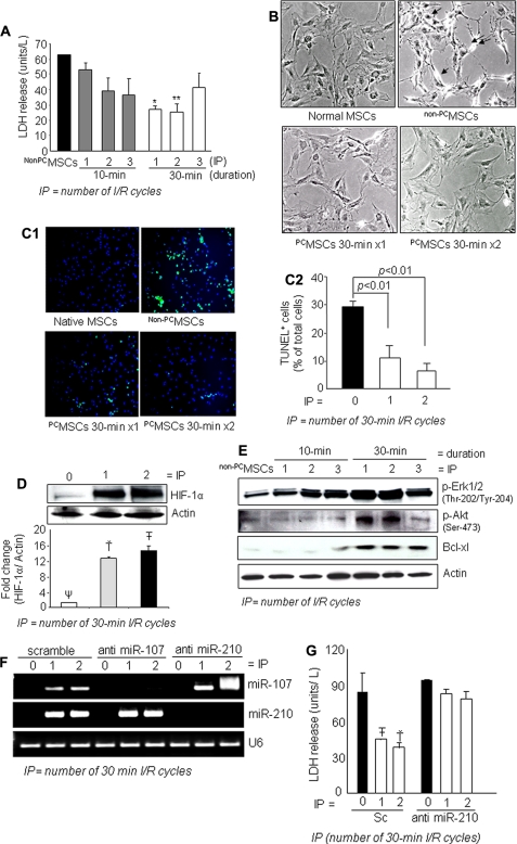
FIGURE 1.
A, IP attenuated cellular injury as examined by LDH release subsequent to 6 h of lethal anoxia. IP with three 30-min cycles was less effective than IP using one 30-min cycle and two 30-min cycles (* and ** versus all other groups of cells, p < 0.05). B, morphological changes of MSCs by 6 h of anoxia included rounding off and shrinkage of cells, which was prevented by IP (magnification, ×200). C1 and C2, representative fluorescence images and quantitative analysis of TUNEL positivity showing decreased number of TUNEL+ cells in PCMSCs (p < 0.01 versus non-PCMSCs) (magnification, ×200; green, TUNEL+ nuclei; blue, DAPI). D, Western blot showing higher HIF-1α in nuclear fraction (two cycles of 30-min I/R) compared with PCMSCs (one cycle of 30-min I/R) and non-PCMSCs. (Ť and T̄ vs. Ψ, p < 0.05). E, Western blot showing higher activation of ERK1/2, Akt, and Bcl.xL in PCMSCs (two cycles of 30-min I/R) compared with PCMSCs (one cycle of 30-min I/R) and non-PCMSCs. F, RT-PCR showing successful and specific knockdown of endogenous miR-210 and miR-107 by transfection with antisense molecules specific for respective miRs. Knock down of miR-107 was used as a control to show the specificity antisense interference. G, abrogation of miR-210 led to loss of cytoprotection by IP in MSCs as determined by LDH release assay. Abrogation of miR-210 completely abolished IP-induced cytoprotection (T̄ and Ť versus all other groups of cells, p < 0.05).

