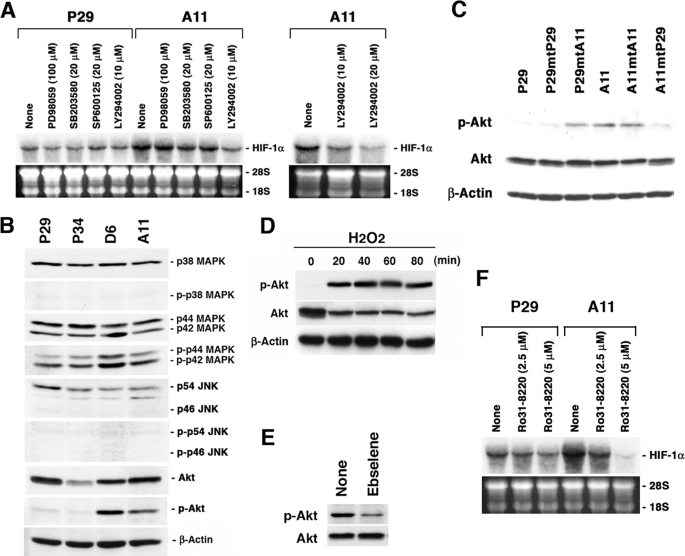
FIGURE 5.
PI3K-Akt and PKC pathways are involved in the ROS-mediated HIF-1α mRNA overexpression. A, P29 and A11 cells were treated with dimethyl sulfoxide, PD98059, SB203580, SP600125, and LY294002 at the indicated concentrations for 18 h. Total RNA was extracted and subjected to Northern blot analysis. The blots were hybridized with a 32P-labeled HIF-1α cDNA. Ethidium bromide staining of the gel is also shown. B, cell lysates prepared from P29, P34, D6, and A11 cells were dissolved by SDS-PAGE. Proteins and phosphorylated proteins and β-actin, which served as a loading control, were detected by immunoblotting. C, cell lysates prepared from P29, A11, and the cybrids were subjected to immunoblotting to detect Akt and phosphorylated Akt. β-Actin served as a loading control. D, P29 cells were treated with 25 μm H2O2 for up to 80 min. Cell lysates were prepared and subjected to immunoblotting as in C. E, A11 cells were treated with ebselene (20 μm) for 18 h. Cell lysates were prepared and subjected to immunoblotting as in C. F, P29 and A11 cells were treated with Ro31-8220 at the indicated concentrations for 18 h. Total RNA was analyzed as in A.

