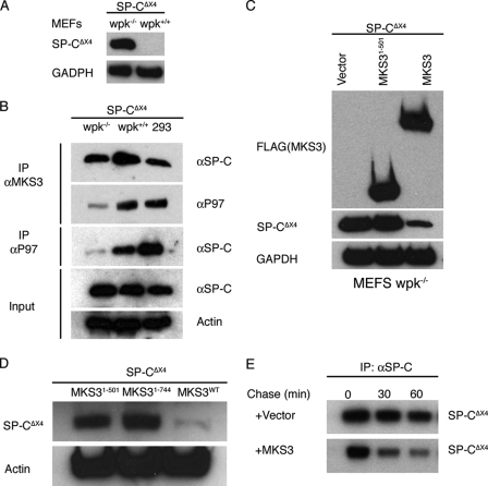
FIGURE 4.
ERAD is decreased in MEFs expressing mutant MKS3. A, MEFs were isolated from day 13 wpk+/+ and wpk−/− embryos and transiently transfected with SP-CΔexon4 (SP-CΔX4). 36 h later, cells were analyzed by SDS-PAGE/Western blotting with anti-SP-C antibody. B, MEFs and HEK293 cells were transiently transfected with SP-CΔexon4. wpk+/+ MEFs and HEK293 cells were treated with MG-132 to promote accumulation of mutant SP-C; all cells were treated with DSP to cross-link interacting proteins. Cell lysates were immunoprecipitated (IP) for endogenous MKS3 or p97 and analyzed by SDS-PAGE/Western blotting with anti-SP-C and anti-p97 antibodies. C, wpk−/− MEFs were transiently cotransfected with SP-CΔexon4 and vector, MKS3-(1–501), or full-length MKS3. 48 h later, cell lysates were analyzed by SDS-PAGE/Western blotting with anti-FLAG, anti-SP-C, and anti-glyceraldehyde-3-phosphate dehydrogenase (GAPDH) antibodies. D, the experiment in C was repeated with MKS3-(1–744). E, wpk−/− MEFs were transiently cotransfected with SP-CΔexon4 and vector or full-length MKS3. 24 h later, cells were labeled with [35S]Met/Cys for 30 min, after which the labeling medium was replaced with chase medium. Cell lysates were immunoprecipitated with anti-SP-C antibody and analyzed by SDS-PAGE/autoradiography.

