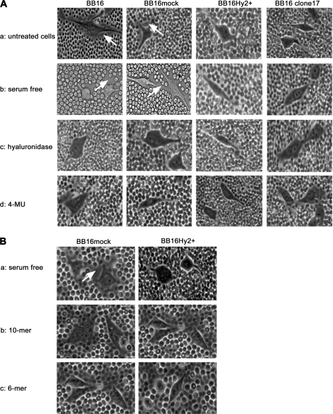
FIGURE 2.
Effect of Hyal2 overexpression and exposure to HA oligosaccharides on the pericellular coat. The latter was visualized by a particle exclusion assay in phase-contrast microscopy; representative images of cells without (BB16 and BB16mock) and with (BB16Hy2+ and clone17) Hyal2 overexpression are shown. Arrows highlight the external border of the coat in each cell type. A, effects of four different conditions: (a) usual culture medium containing 10% fetal bovine serum; (b) the same medium after 16-h serum deprivation; (c) following 1-h treatment with 5 units/ml Streptomyces hyaluronidase in serum-free medium; (d) following 16-h treatment with 100 μm 4-methylumbelliferone (4-MU) in serum-free medium. B, effects of: (a) control serum-free medium; and addition of (b) 10-mer or (c) 6-mer HA oligosaccharides at 100 μg/ml for 16 h in serum-free medium.

