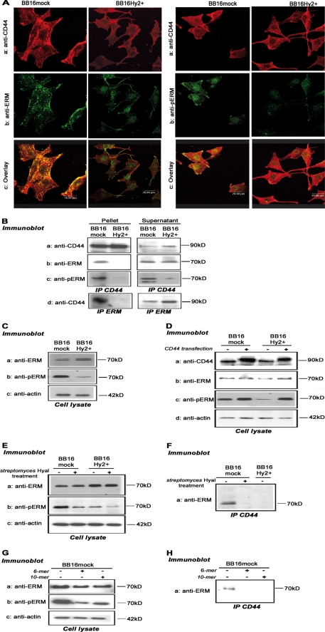
FIGURE 6.
The Hyal2-CD44-ERM interaction. A, representative confocal microscopic images of immunofluorescent CD44 (red, OX49 antibody), ERM, or phosphorylated ERM (pERM, green), and their co-localization (yellow in the overlay images). B, co-immunoprecipitation of CD44 (OX49 antibody) and ERM with each other and with pERM. Supernatants of the antibody-antigen-beads complex are shown as controls. C, Western blots of ERM, pERM, and actin in cell lysates (5 μg of protein). D, the same blots, as well as CD44 detection, were repeated 24 h after transfection of a rat CD44 cDNA and compared with untransfected controls. E and F, Western blots of ERM, pERM, and actin in cell lysates, and detection of pERM in CD44 immunoprecipitates, following 1-h treatment with 5 units/ml Streptomyces hyaluronidase or vehicle. G and H, same detection steps following 16-h treatment with 100 μg/ml 6-mer and 10-mer HA oligosaccharides.

