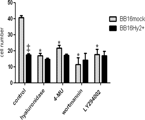
FIGURE 7.
Wound healing assays. Cells were grown to confluency, then induced to re-populate a wound created by a sterile razor blade. To prevent growth during migration, cells were pre-treated with 4.5 mm mitomycin C for 2 h. After band-stripping, they were allowed to migrate into the wound for 6 h in medium containing 10% fetal calf serum alone or supplemented with 5 units/ml Streptomyces hyaluronidase, a HA synthase inhibitor (4-MU at 100 μm), or PI3K inhibitors (50 nm wortmannin or 5 μm LY294002). Results are presented as means ± S.E. of the number of cells that had colonized the wound margin in random microscopic fields (*, p < 0.001 versus untreated BB16mock cells, n = 5 in each group; ‡, p < 0.001 versus BB16mock cells, n = 5).

