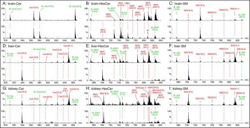
FIGURE 3.
Precursor ion mass spectra of sphingolipids from control (upper spectrum) and Cers2gt/gt (lower spectrum) mice. Ceramides (Cer) (A, D, and G) and monohexosylceramides (MHC) (B, E, and F) were detected with the precursor ion scan m/z +264 specific for sphingolipids with d18:1-sphingosine. Sphingomyelins (SM) (C, F, and I) were analyzed with the precursor ion scan m/z +184 specific for the phosphorylcholine headgroup. Fatty acid chain lengths of SM were interpreted with the main sphingoid base sphingosine (d18:1) present in the analyzed tissues. A–C, brain; D–F, liver; G–I, kidney. Internal standards are in green, and endogenous sphingolipids are in red. m/z 808.5 marked with an asterisk in the lower spectrum of B is no hexosylceramide (HexCer), as verified with a second hexosylceramide-specific scan, neutral loss +180 atomic mass units. Quantitative evaluations of the mass spectra include also the standards Cer and hexosylceramide as well as SM standards with C31:0 acyl moieties and are shown in Fig. 4.

