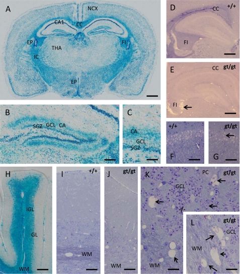
FIGURE 6.
lacZ reporter gene expression and phenotypic abnormalities in the nervous system of cers2gt/gt mice. A, within the brain, β-galactosidase is expressed in all neuronal strata of the neocortex (NCX) and the white matter of the internal capsule (IC), corpus callosum (CC), and fimbria hippocampi (FI). In contrast, neuronal stain in the thalamus (THA) appears much weaker but is still present. Note the especially strong β-galactosidase staining within the CA1 field of the cornu ammonis and the ventricular ependyma (EP). B and C, within the dentate gyrus, β-galactosidase staining is especially intense within the subgranular zone (SGZ) containing the life-long active neuronal stem cell reservoir for the granule cell layer (GCL). Interestingly, the β-galactosidase signal also occupies the sublayer for commissural and associational afferents (CA) within the dentate molecular zone, an effect to be ascribed to an as yet not understood compartmentalization of β-galactosidase. D and E, when glycid ether-embedded and toluidine blue/pyronin G-stained samples of the telencephalon are compared with wild type mice (D), it is striking that the stainability of the white matter (corpus callosum and fimbria hippocampi) for basic dyes like toluidine blue is lost in cers2gt/gt mice (E), pointing toward severe biochemical alterations in myelin sheaths encompassing the loss of acidic compounds. Note the shrunken and ragged appearance of the fimbria in cers2gt/gt mice pointing toward severe axon loss (arrow in E). F and G, despite the high expression of the lacZ reporter in the hippocampal field CA1, only limited irregularities in the cell density of the pyramidal cell layer could be found (arrow in G). H, in the cerebellum, β-galactosidase is equally highly expressed in both the white matter (WM) of the medullary tree as well as in the internal granular layer (IGL), whereas the signal in the ganglionar layer (GL) is relatively weak and may reside rather in the Bergmann glia cell bodies than in the Purkinje cells. I and J, when cerebella of wild type (I) and cers2gt/gt (J) mice are compared, it is apparent that, similar to the telencephalon, the stainability of the white matter is largely lost. Again, this is not due to the loss of myelin sheath because they can be still discerned at low contrast but appears to be due to an alteration of their composition. K and L, an additional effect of CERS2 deficiency in the cerebellum, not seen in the telencephalon, is the formation of numerous small cysts (arrows) both within the gray matter of the internal granular layer (K) and the white matter (L), indicating the loss of both myelinated axons and granular layer interneurons. Calibration bars, 1 mm in A, 200 μm in B, 50 μm in C, 500 μm in D and E, 75 μm in F and G, 150 μm in H, 50 μm in I and J, and 20 μm in K and L.

