FIGURE 2.
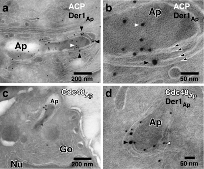
The ERAD components Der1Ap and Cdc48Ap localize to the membrane compartment surrounding the apicoplast. Host cells infected with a transgenic T. gondii line expressing an HA-tagged version of Der1Ap were fixed, frozen, and sectioned with a cryo-ultramicrotome. Sections were incubated with a rat antibody to HA (a, b, d, black arrowheads), a rabbit serum to ACP (9) (a and b, white arrowhead), and a newly developed rabbit antibody against recombinant Cdc48Ap (c and d, white arrowhead with black outline, see also supplemental Fig. S1) followed by secondary antibody conjugated to 12 or 18 nm colloidal gold, stained with uranyl acetate/methylcellulose, and analyzed by transmission electron microscopy. Panel b shows a higher magnification of the cell shown in panel a, the four membranes of the apicoplast are indicated by black arrowheads outlined in white. Ap, apicoplast; Go, golgi; Nu, nucleus.

