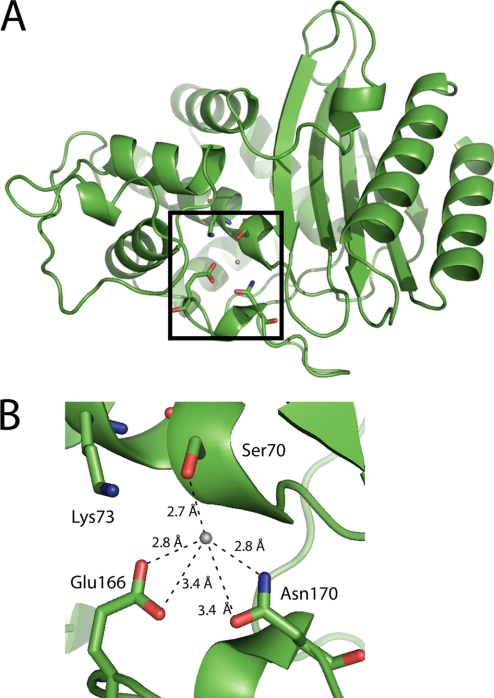
FIGURE 3.
The diagram illustrates the overall structure and the active site of the wild-type TEM-1 enzyme (22). A, a cartoon representation of the overall structure of TEM-1 β-lactamase is shown with the active site boxed. B, the active site is shown with several of the conserved residues labeled and the hydrogen bonding environment near the hydrolytic water illustrated including Asn170, which is one of the three residues that position the water (Protein Data Bank code 1BTL) (22).

