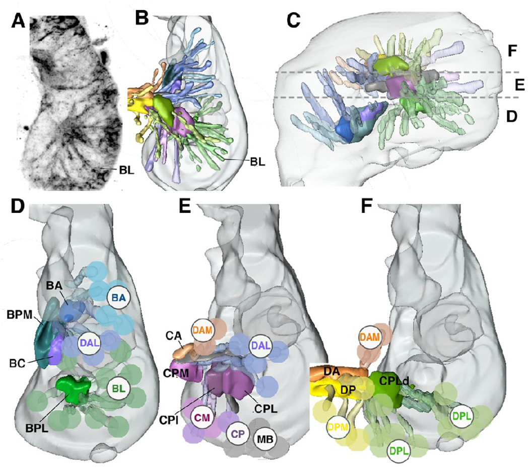
Fig.1.
Projection of groups of neural lineages of the Drosophila central brain.
A: Z-projection of horizontal confocal sections of a late stage 15 brain hemisphere, labeled with UAS-synaptobrevin-GFP driven by elav-Gal4 which visualizes primary lineages. Medial is to the left, anterior down. Z-projection was generated by combining ten contiguous focal planes taken at 1µm increment; it represents a “slice” of the brain containing the ventral lineages and compartments. Lineages can be recognized by characteristically positioned primary axon tracts; one tract, formed by one of the BL lineages, is shown as example. B, C: 3D digital models of late embryonic brain hemisphere; B presents dorsal view (anterior to the top), E shows lateral view (anterior to the left). Brain is outlined in gray. Primary axon tracts of groups of primary lineages are rendered in different colors. Emerging compartments are rendered in the same colors as the groups of lineages that most strongly contribute to (scaffold) the corresponding compartment. The same color key is applied throughout panels B–F. Horizontal hatched lines in C demarcate boundaries between the dorsal cerebrum (F), middle cerebrum (E), and ventral cerebrum (D). Groups of lineages located at these levels are shown separately in a dorsal view in the models presented in panels D–F. Here, aside from the primary axon tract, a lineage is schematically represented by a colored sphere and identified by acronyms (BA baso-anterior; BL baso-lateral; CM centro-medial; CP centro-posterior; DAM dorso-anterior medial; DAL dorso-anterior lateral; DPL dorso-posterior lateral; DPM dorso-posterior medial; MB mushroom body; for detail, see Younossi-Hartenstein et al., 2006). Abbreviations of compartments are identified by black capital letter without sphere: BA baso-anterior; BC baso-central; BPL baso-posterior lateral; BPM baso-posterior medial; CA centro-anterior; CPI centro-posterior intermediate; CPL centro-posterior lateral; CPLd dorsal domain of CPLd; CPM centro-posterior medial; DA dorso-anterior; DP dorso-posterior. Note that in this and the following figures, lineages of th tritocerebrum, which have been insufficiently mapped up to the present point, are not shown.
Bar: 20µm

