Fig.3.
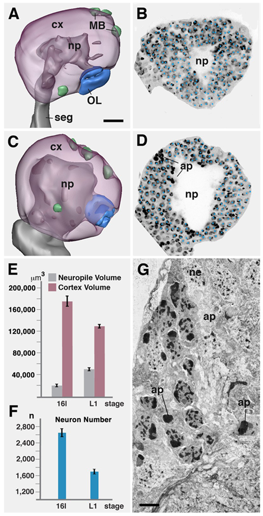
Apoptotic cell death of primary neurons at the embryo-larva transition. A, B: stage 16 embryonic brain hemisphere. A shows 3D digital model (lateral view, anterior to the left) in which neuropile (np) is rendered grey, cortex (cx) transparent magenta. Active neuroblasts (green) are associated with the mushroom body (MB) and basal-anterior compartment. Optic lobe primordium (OL) is rendered blue. B shows a parasagittal confocal section in which nuclei of neurons and glial cells were labeled with Sytox (individually marked by blue dots). C, D: Representation of first instar larval brain hemisphere as digital 3D model (C) and confocal section (D; same scale as A and B). Note increase in diameter of neuropile, and concomitant decrease in thickness of cortex. Dense bodies in D (ap) are pycnotic nuclei of apoptotic neurons/glial cells. E: Graph comparing volume of neuropile and cortex between stage 16 embryo (left bars) and first instar larva (right bars; both hemispheres combined). F: Graph comparing number of cells in cortex of one brain hemisphere between stage 16 embryo (left) and first instar larva (right). G: Electron micrograph of section of first instar brain cortex, showing somata of neurons (ne) and apoptotic nuclei (ap).
Bars: 10µm (A); 2.5µm (G)

