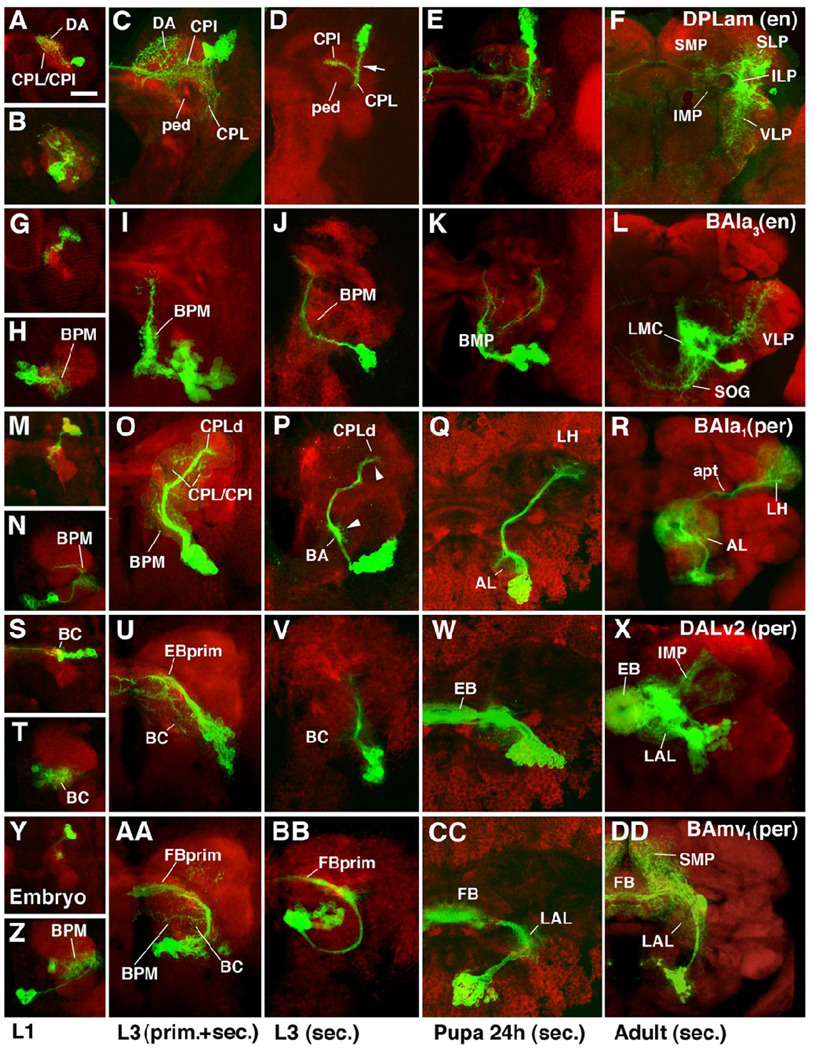
Fig.5.
Morphogenesis of selected lineages marked by specific Gal4 driver lines. Panels represent Z projections of confocal sections of brain hemisphere labeled with anti-DNcadherin (red; neuropile) and antiGFP to visualize individual lineages in which UAS-mCD8-GFP is driven by driver line engrailed-Gal4 (A–L) or period-Gal4 (M-DD). Panels are organized such that each row shows one specific lineage at four different developmental stages, indicated at bottom of figure. The first panel (e.g., A) shows embryo, the second (e.g., B) first instar larva; the third (e.g., C) late third instar in which both primary and secondary neurons are labeled; the fourth (e.g., D) late third instar with labeling of only secondary neurons; the fifth (e.g., E) pupa 24h after puparium formation; the sixth (e.g., F) adult. Except for panels depicting embryos, which present dorsal view, all panels show anterior view. A–F: Engrailed–positive DPLam lineage; type C. Primary neurons arborize widely in CPL, CPI, DP/DA, and CA compartments (A–C). Secondary axon tract (arrow in D) follows primary neurons into CPL compartment; here it branches into medial branch that crosses over peduncle (ped) into CPI, and a ventral branch remaining in CPL. Branches of secondary neurons develop along these axes (E, F), filling inferior medial and inferior lateral protocerebrum (IMP, ILP; descendants of CPI and CPL, respectively). Peripherally, branches continue into neighboring regions of superior lateral protocerebrum (SLP) and ventro-lateral protocerebrum (VLP). Interestingly, there seems to be no arborization of secondary neurons in superior medial protocerebrum (SMP), the descendant of the DA compartment that was filled with primary neurites of the DPLam lineage (compare panels C and F). G–L: Engrailed-positive BAla3 lineage. Both primary and secondary components of this lineage project long undivided axon tract posteriorly (D lineage; see Fig.4) and branch in BPM compartment. In addition, some secondary branches project latero-dorsally into VLP, and ventro-medially into subesophageal ganglion (SOG). M–R: Period-positive BAla1 lineage; type PD. Primary neurons project to BPM compartment and the region around the peduncle (CPI, CPL). Secondary neurons represent the class of multiglomerular antennal projection neurons. They form proximal arborizations (dendrites) in antennal lobe (AL in Q and R), and axonal arborizations in lateral horn (LH). Note how proximal and distal arbors are prefigured by tufts of filopodia along secondary axon tract in larva (arrowheads in P). S–X: Period-positive DALv2 lineage; type PD. Primary arborizations in BC compartment and the primordium of the ellipsoid body (EBprim), which in larva forms part of CPM compartment. Secondary neurons of this lineage form majority of ring neurons of ellipsoid body (EB). Other neurons of the same lineage form additional arborizations in lateral accessory lobe (LAL; descendant of BC compartment) and inferior medial protocerebrum (IMP). Y-DD: Period-positive BAmv1 lineage. D lineage with conspicuous crescent-shaped tract, projecting first posteriorly, then dorsolaterally, and finally dorso-medially towards the primordium of the fan-shaped body (FBprim), which forms part of the CPM compartment of the larval brain. Arborizations in BC, BPM and FBprim. Secondary axons follow the same trajectory and branch in LAL, fan-shaped body (FB), and superior medial protocerebrum (SMP).
Bar: 20µm

