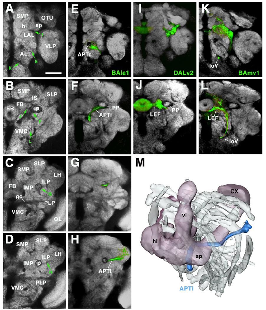
Fig.7.
Secondary axon tracts develop into long fiber bundles of adult brain. A–L show frontal confocal sections of adult brain hemisphere labeled with antibody Nc82 (neuropile; white). Rows correspond to the antero-posterior level of brain slice shown. Top row (A, E, I, K) corresponds to anterior level, showing optic tubercle (OTU) and horizontal lobes of mushroom body (hl). Slices shown in second row (B, F, J, L) are slightly more posteriorly, at level of ellipsoid body (EB). Third row taken at level of fan-shaped body (FB), bottom row at level of lateral horn (LH). In panels of left column (A–D) Nc82 was only marker used. In panels of second column (E–H), the secondary neurons of the BAla1 lineage are labeled by GFP (driven by period-Gal4). Third column (I, J) shows period-positive DALv2 lineage; fourth column (K, L) shows period-positive BAmv1 lineage. Note that the coherent secondary axon tracts of lineages now form long fiber bundles which are visible as Nc82-negative (i.e., synapse-free) “tunnels”, indicated by green arrows in panels of left column. Axons of BAla1 join the common antenno-protocerebral tract (APTc; E), a massive, Nc82-negative landmark. At the level of the ellipsoid body (F) BAla1 axons diverge laterally and then pass underneath the peduncle (p; in F and G), forming the medio-lateral APT (APTl). Further posteriorly, this tract continues straight towards the lateral horn (D, H). Axons of DALv2 (I, J) pass underneath the horizontal lobe of the mushroom body (I; see arrow in A) and then continue medially towards the ellipsoid body (J, B), forming the lateral ellipsoid fascicle (LEF). BAmv1 axons grow posteriorly as part of the longitudinal ventral fascicle (loV; panels K, L, C, D). Subsequently they turn upward laterally, then medially, to join the LEF at its posterior face (L). M: 3D digital model of left hemisphere of adult brain, shown in antero-dorso-lateral view. Outlines of all long fascicles that produce nc82-negative impressions of more than 1.5µm diameter are shown shaded grey. One bundle, the APTl (marked by the BAla1 lineage; see panels E–H) is shown in color (blue). The mushroom body is shown for orientation (CX calyx; hl horizontal lobes; vl vertical lobes; sp spur). Other abbreviations:
AL antennal lobe; gc great commissure; ILP inferior lateral protocerebrum; IMP inferior medial protocerebrum; LAL lateral accessory lobe; LH lateral horn; OL optic lobe; OTU optic tubercle; PLP postero-lateral protocerebrum; SLP superior lateral protocerebrum; SMP superior medial protocerebrum; VLP ventro-lateral protocerebrum; VMC ventro-medial cerebrum
Bar: 40µm

