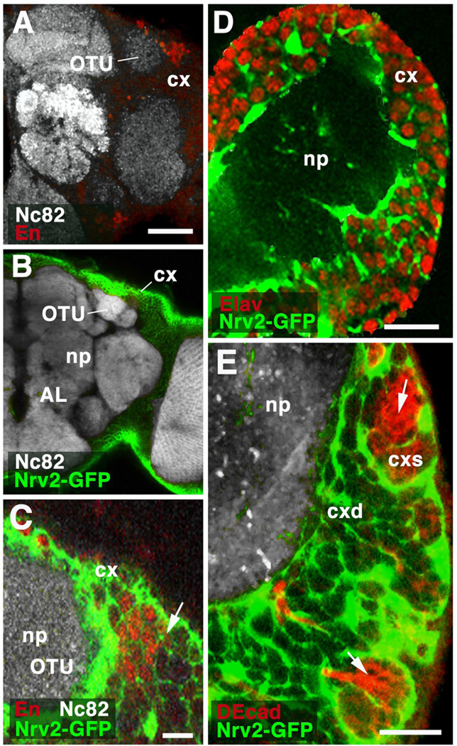
Fig.8.
Relationship of glia and lineages. A–C: Z-projections of frontal sections of adult brain hemisphere, labeled with antibody Nc82 (neuropile; np). In A, anti-Engrailed shows cell bodies of DPLam lineage, located in cortex (cx) flanking the optic tubercle (OTU). In B and C, cortex glia is labeled by GFP driven by Nrv2-Gal4. C presents a high magnification view of the dorso-lateral cortex containing the Engrailed-positive DPLam neurons (red). Cortex glia (green) forms sheaths around each individual neuron. No specialized glial layer demarcates the boundary (arrow) between labeled neurons of DPLam lineage and their unlabeled neighbors, which belong to different lineages. D: Z-projection of frontal section of first instar brain hemisphere. Neuronal nuclei are labeled by anti-Elav; cortex glia is labeled by Nrv2-Gal4>UAS-GFP. At this stage, all neurons in brain cortex (cx) are differentiated, primary neurons. As in adult (see C), each individual differentiated neuron is surrounded by cortex glial lamella. E: Z-projection of frontal confocal sections of late third instar brain hemisphere. As in B – D, cortex glia is labeled (green). Newly born ganglion mother cells and secondary neurons, located in superficial cortex (cxs), are labeled by anti-DEcadherin (red). Note that these undifferentiated secondary neurons form clusters which are enclosed in large glial chambers (arrow). As secondary neurons mature in deep cortex (cxd), they become individually enclosed by glial lamella. Other abbreviations: AL antennal lobe; cxd deep cortex
Bars: 40µm (A, B); 10µm (C, D, E)

