
Figure 6. Basophils release antimicrobial factors upon IgD cross-linking.
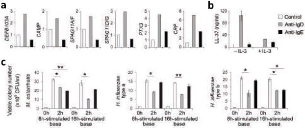
(a) QRT-PCR of DEFB103A, CAMP, SPAG11A/F, and SPAG11D/G transcripts from basophils exposed to microbeads alone (control, open bar), microbead-bound monoclonal anti-IgD (gray bar), or microbead-bound monoclonal anti-IgE (black bar) for 6 h. PTX3 and CRP transcripts were measured after 16 h. mRNAs were normalized to ACTB mRNA. (b) ELISA of LL-37 from basophils stimulated as in a in the presence or absence of IL-3 for 8 h. (c) Growth of Moraxella catarrhalis and Haemophilus influenzae type-a and type-b upon 2-h exposure to culture supernatants from basophils stimulated as in a for 8 h or 16 h. CFU, colony forming unit. Panels a-c summarize 3 experiments (bars indicate s.e.m.; *, p < 0.03; **, p < 0.02; ***, p < 0.01).