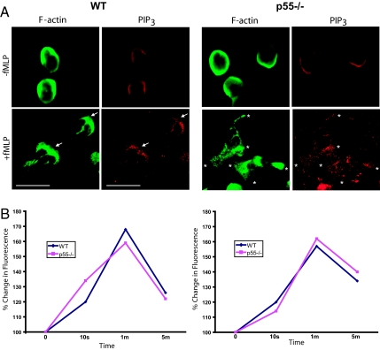
Fig. 4.
PIP3 localization and its content. (A) Localization of PIP3 in p55−/− neutrophils. A monoclonal anti-PIP3 antibody was used to stain PIP3 in resting and activated WT and p55−/− neutrophils. In resting cells, there was a faint signal of PIP3 around the peripheral membrane. Upon stimulation with fMLP, WT neutrophils polarized by accumulating PIP3 toward the leading edge membrane marked by F-actin staining. Upon stimulation, p55−/− neutrophils formed extensions but PIP3 is diffusely localized throughout the cytosol. (B) Neutrophils were incubated with anti-PIP3 antibody and FACS analysis was performed to measure total PIP3 levels after fMLP stimulation. No difference in total PIP3 was observed between WT and p55−/− neutrophils. Two independent experiments are shown.

