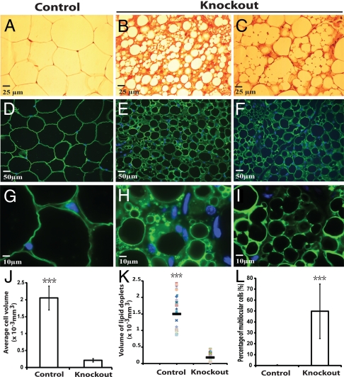
Fig. 2.
Histological and immunofluorescence analysis of gonadal WAT from control and atg7 conditional knockout mice. (A–C) Representative microscopic pictures of H&E stained sections of uterine WAT from control (atg7flox/flox, A) and adipose-specific atg7 conditional knockout mice (atg7flox/flox; aP2-Cre, B and C). (D–I) Representative microscopic pictures of immunofluorescence assays of uterine WAT from control (D and G) and atg7 conditional knockout mice (E, F, H, and I) with Perilipin A antibody. D–F were pictures of low magnification and G–I were pictures of high magnification. (J–L) Quantification of average cell volume, lipid droplet volume, and percentage of multilocular cells, as indicated, of uterine WAT from control and atg7 conditional knockout mice. ***, P < 0.001, Student's t test. The data show representative results of tissues from six pairs of female mice (control and atg7 knockout).

