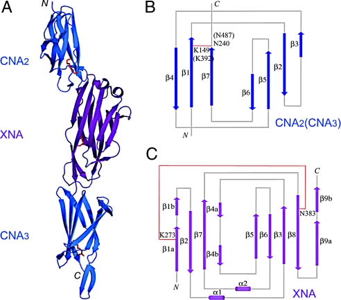
Fig. 1.
Crystal structure of BcpA* and topology diagrams of the CNA and XNA domains. (A) The crystal structure of BcpA* was solved using MAD. The CNA2 and CNA3 domains are shown as blue ribbons and the XNA domain purple. The Lys and Asn residues forming the intramolecular amide bonds in each of the 3 crystallized domains are shown as red sticks. Topology diagrams of the CNA (B) and XNA (C) domains show the arrangement of β-strands in the reverse Ig-fold (CNA) and jelly-roll fold (XNA). Both CNA2 and CNA3 share the same overall topology. β-strands are shown as arrows and α-helices as cylinders. Intramolecular amide bonds are shown as red lines.

