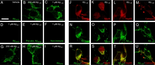
Fig. 1.
Confocal images of Aβ, MAP2, NeuN, Hsp60, and calnexin in cortical neurons after exposure to Aβ-related peptides. Cells were exposed to the indicated concentration [or 1μM (J−U)] of human Aβ1−40, Aβ1−42, reversed Aβ (Aβ40−1 and Aβ42−1), or vehicle for 60 min, fixed in 3% PFA, and stained by [anti-human Aβ (clone 4G8) (A–D, G−I, N, and R), anti-Aβ1–40 (O–P and S–U), preabsorbed anti-Aβ (clone 4G8) (E) or non-immune IgG (F)]/Alexa Fluor 488 anti-IgG (green), anti-MAP2/Alexa Fluor 568 anti-IgG (red) (J and R) and [anti-NeuN (K and S), anti-Hsp 60 (L and T), or anti-calnexin (M and U)]/Alexa Fluor 546 anti-IgG (red). Scale bar, 10 μm. Hoechst 33342 staining and phase contrast images of the same field of cells in panels of A, C, H, D, or Fig. S4 A–D are represented in Fig. S4 E–H and M–P, respectively.

