Fig. 3.
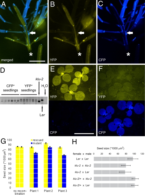
KLU acts locally in the maternal tissue of developing flowers to promote seed growth. (A–C) Fluorescence micrographs of one of the three genetically grafted plants analyzed in G, showing the transition in the inflorescence from genotypically WT tissue marked by YFP fluorescence (yellow) to klu-2 mutant tissue marked by CFP expression (blue). The approximate point of transition is indicated by the solid arrow. The asterisk indicates a section of stem covered by the shadow of the silique at the bottom of the image. (A) Merged image. (B) YFP channel. (C) CFP channel. (D–F) Analysis of seedlings germinated from seeds that were harvested from WT (E) or mutant (F) siliques of the plant shown in A–C. (D) PCR analysis of seedlings shown in E and F to detect the presence of the WT KLU allele in the rescue construct. Whereas YFP-positive seedlings still contain the WT allele, it has been lost from the CFP-positive but YFP-negative seedlings. Amplifications on nontransgenic klu-2 mutant and Ler WT DNA are shown as controls. (E) Seedlings from YFP-positive WT silique. (F) Seedlings from CFP-positive klu-2 mutant silique. (E and F, Top) YFP channel. (E and F, Bottom) CFP channel. (G) Size of seeds from WT (yellow bars) and mutant (blue bars) siliques of three independent chimeras. The leftmost two bars show seed size from early (Left, flowers 1–6) and late (Right, flowers 20–25) siliques of a nonrecombined rescue plant for comparison. In all three recombined plants, seeds from mutant siliques were significantly smaller at P < 0.05 (two-tailed t test). (H) Size of seeds resulting from the indicated reciprocal crosses. Note that error bars in H show SD as a measure of variability in the seed populations. Values shown are mean ± SEM (G) and mean ± SD (H). (Scale bars: 5 mm.)

