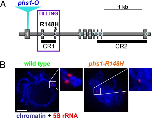
Fig. 4.
The maize phs1-R148H mutation. (A) The maize Phs1 gene. Blue triangle = position of Mutator transposon insertion resulting in the original phs1-O mutation. Violet box = region used for the TILLING screen. Asterisk = positions of the phs1-R148H mutation. (B) Paring of 5S rRNA loci in a wild-type and phs1-R148H mutant meiocytes. Both loci in the mutant are associated with nonhomologous chromosome segments. Closeups of homologously paired 5S rRNA loci in the wild-type and one of the loci in the mutant are shown in insets. The other 5S locus in the mutant is visible in the lower right corner of the cell. Images are flat projection of several consecutive optical sections but do not represent entire nuclei. (Scale bar, 5 μm.)

