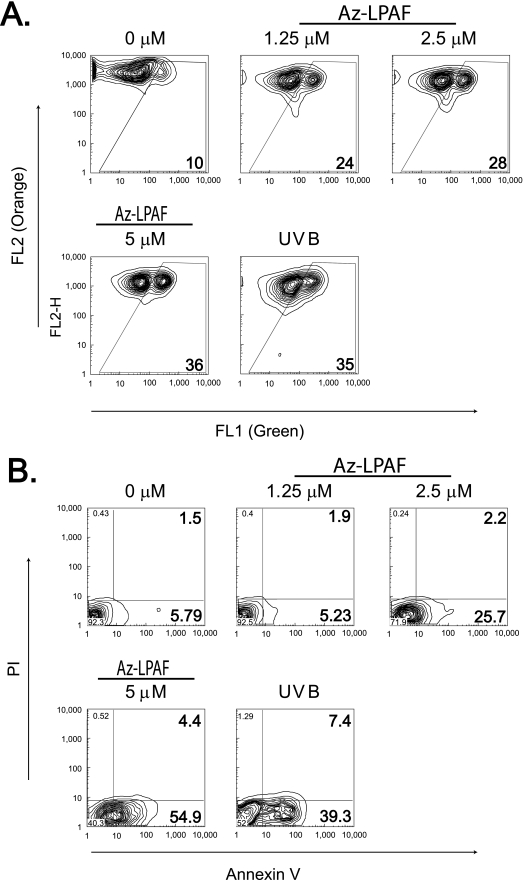
FIGURE 1.
The oxidatively truncated phospholipid Az-LPAF depolarized mitochondria and initiated apoptosis in intact HL-60 cells. A, mitochondrial transmembrane potential is compromised in intact HL-60 cells by Az-LPAF. HL-60 cells were incubated with the stated concentrations of Az-LPAF for 4 h, and then stained with the fluorescent potentiometric dye JC-1 as described under “Experimental Procedures.” The positive control was irradiation with 400 J/m2 UVB for 5 min. Two-color flow cytometry in the green FL1 (monomeric dye) and red FL2 channel (mitochondrial aggregates) are presented as topologic representations (n = 3). B, Az-LPAF initiates early apoptotic events. HL-60 cells were incubated in RPMI with the stated concentrations of Az-LPAF for 6 h in media, or were irradiated with UVB. The washed cells were stained with Alexa 488-conjugated annexin V and propidium iodide to stain nuclei of cells with failed permeability barriers as defined under “Experimental Procedures” (n = 3).

