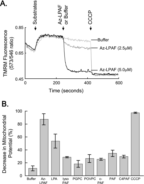
FIGURE 2.
Isolated mitochondria exposed to truncated phospholipids rapidly lose their transmembrane potential. A, Az-LPAF rapidly depolarized isolated mitochondria. Mitochondria isolated from rat liver were loaded with the potentiometric dye TMRM as described under “Experimental Procedures,” and then the ratio of fluorescence intensity from excitation at 573 and 546 nm with emission at 590 nm was recorded as a function of time. The oxidizable substrates glutamate and malate were added (first arrow), and then the stated concentration of Az-LPAF or buffer was added (second arrow). Fluorescence by mitochondria in a completely depolarized state was determined by the addition of the protonophore CCCP (5 μm) at the third arrow (n = 5). B, Az-LPAF is the most effective, but not the only, phospholipid to depolarize mitochondria. Isolated mitochondria labeled with TMRM were treated with 2.5 μm of various short-chain phospholipids or lysolipids, and the change in TMRM fluorescence after 200 s of exposure relative to the loss of membrane potential after exposure to CCCP was recorded. The data from three independent experiments are shown with standard error (n = 3). LPA, lysophosphatidic acid; PGPC, palmitoyl glutaroyl phosphatidylcholine; POVPC, palmitoyl oxovaleroyl phosphatidylcholine; c-PAF, carbamoyl platelet-activating factor; C4-PAF; butyroyl platelet-activating factor.

